Tiny Health Bacterial Toxins Frequently Asked Questions

A phone showing Toxin genes detected in a Tiny Health gut health sample

Summary

.
.

Below, you'll find answers to common questions about bacterial toxins. This guide will help you understand bacterial toxins, how they impact your gut health, and how the Tiny Health Gut Test can identify them. To access this feature, log in to your account to explore your existing gut health report or purchase a Gut Health Test. 

What are bacterial toxins?

Bacterial toxins are like tiny weapons that cause illness. They can target different parts of our body and mess up normal functions. For example, some toxins attack our cells, causing cell death. Others impact how the systems in our bodies talk to each other, leading to inflammation and other health issues [1]. Our gut microbiome plays a role in this, too. Most gut bacteria are helpful. But some can make toxins that can cause digestive issues and affect our health. 

Does the Tiny Health gut test detect actual bacterial toxins in the gut?

While the Tiny Health Gut Test doesn’t show the toxins made by bacteria in a direct way, it identifies genes in the bacteria that have these toxins that we call “bacterial toxin genes”. Each bacterium carries a unique set of genes, including some that make toxins. By looking at the microbial DNA in your gut, the test can find out if your gut bacteria might produce harmful toxins.

Where do I find bacterial toxins in my report?

In the ‘Disruptive microbes’ category of your Gut Health Test report, you’ll find the section Opportunistic Pathogens. In this list, some species may highlight toxins detected. Click any of them to view the details of the toxins. 

My sample is positive for bacterial toxin genes. What does this mean?

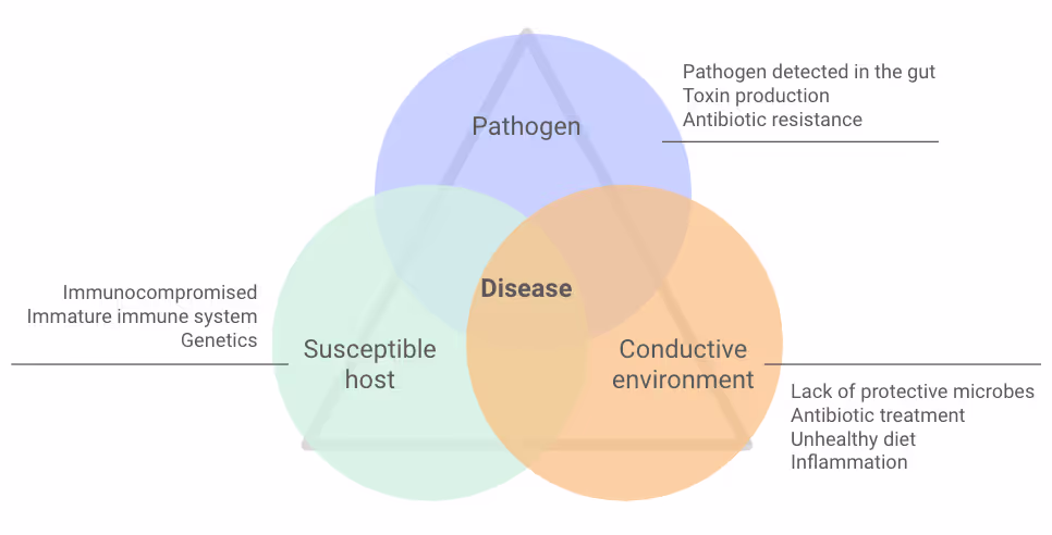
Certain bacteria in your gut can potentially produce toxins. But it's important to know that if your sample is positive, it doesn’t mean toxins are active or causing harm now. Their presence shows that these bacteria could produce toxins under certain conditions. Your diet, lifestyle, immune system, and gut health help influence if these bacteria become active, make toxins, and cause illness (Figure 1).

Figure 1. For disease to occur, health issues, a weakened gut, and harmful germs must all be present at the same time.

What does it mean if my sample doesn't have all the genes for a toxin?

Having only some genes for a toxin means the bacterium doesn't have all the instructions needed for the toxin to work. Bacterial toxins often have many parts that work together to harm the body. If your results show only some of the toxin genes, it means the bacteria in your gut might be able to make part of the toxin.

In other words, these bacteria might not make you as ill as ones with all the toxin genes.

My results show an opportunistic pathogen, but bacterial toxin genes were not detected. What does that mean?

Some strains of the same bacterial species can have different toxin genes. This means that while specific strains can make toxins, others might not have these genes at all or have different versions of them.

Sometimes, toxin genes can live on DNA that bacteria can pass around or lose over time.

Results are often different when opportunistic pathogens don’t have toxin genes. If you get an infection, it could be less severe than one with toxin genes. Without toxin genes, pathogens can't do as much harm. 

It's also possible that the toxin genes are there, but in such small amounts that they aren’t detected.

Can having toxin genes in my gut predict health outcomes?

Sometimes. Having toxin genes can increase the risk for certain conditions. But it doesn’t mean you'll get them.

For example, if you have Clostridioides difficile (C. diff) in your gut, you might get diarrhea after taking antibiotics. If you test positive for C. diff’s toxin A and B gene, it means the C. diff can cause severe antibiotic-related diarrhea. If this happens, supporting your gut during and after antibiotics is important.

In short, having a bacterial toxin gene doesn't always mean you will get a specific health problem. But it shows that some opportunistic pathogens in your gut might be able to cause disease.

What can I do to reduce the levels of bacterial toxin genes in my gut microbiome?

To lower the levels of bacterial toxin genes in your gut, you need to target specific opportunistic pathogens that carry them. 

When you balance your gut bacteria, it helps to reduce the harmful germs that carry these toxin genes. If your levels are high, your Action Plan will likely recommend that you:

  • Eat a balanced diet rich in fiber
  • Avoid processed foods and sugars
  • Take a targeted probiotic supplement

What bacterial toxin genes do Tiny Health gut tests identify?

Below is the list of toxin genes that may show up in the Tiny Health Gut Test.

Staphylococcus

Exfoliative toxin type A attacks desmoglein-1, a key protein that helps skin cells stick together. When the cells disconnect, blisters occur [2]. The toxin's role in the gut is unknown because it targets skin cells. But, if the gut has Staphylococcus, it might increase the risk of skin infections [3].  

Staphylococcal enterotoxins cause food poisoning. Staphylococcal enterotoxins cause a strong inflammatory response and help the infection spread [4]. After eating contaminated food, severe diarrhea, nausea, and stomach cramps may occur [5]. They can also cause toxic shock syndrome. These toxins are often found in children’s guts [6], [7].

Toxic shock syndrome toxin 1 makes immune cells release substances that cause inflammation, fever, and shock [8].

Leukocidins are toxins that make holes in the surface of immune cells and red blood cells. They disrupt cell balance and lead to inflammatory cell death [9].

Superantigen-like protein 1, Superantigen-like protein 5/11, Superantigen-like protein 7 are harmful Staphylococcus aureus toxins. They change how immune cells work and allow the bacteria to survive and stay in the body [10], [11].

Streptococcus

C3 family ADP-ribosyltransferase disrupts the inside of immune cells and makes the bacteria more harmful [12], [13], [14]. 

cAMP factor forms bubbles inside macrophages, making it harder for them to destroy pathogens or foreign particles [15], [16]. This helps the bacteria stick to and enter cells from the throat.

Streptococcus agalactiae (Group B Strep)

CylE protein is an enzyme that helps make a toxic pigment from Group B Strep. The pigment ornithine rhamnolipid harms red blood cells, platelets, and other immune cells [17], [18].

Pseudomonas aeruginosa

Adenylate cyclase disrupts immune responses and helps bacteria grow, making P. aeruginosa more harmful [19], [20].

Exoenzyme S messes up how macrophages move and change shape. This makes it harder for them to crawl around and swallow things [21]. It also stops epithelial cells from making DNA. This disrupts their structure and interferes with how they talk to each other [22].

Exotoxin A is the most harmful toxin from P. aeruginosa. It stops cells from making proteins, causing them to die [23].

Exoenzyme U interferes with cell shape and disrupts cellular barriers. It also causes tissue swelling [24].

Escherichia coli

Cytolethal distending toxin, subunits A, B, and C makes cells swell, damages DNA, and leads to their death. It’s also linked to causing long-lasting infections [25]. Since it destroys DNA, scientists think it may have a role in cancer promotion [26]. Escherichia coli bacteria that produce this toxin have been found in people with colon cancer [27]. This toxin has only been tested on animals, so we need more research to understand its effects on the gut. All subunits need to be present to have toxic effects.

Shiga toxin, subunits A and B stop cells from making proteins. Subunit A breaks the cell’s protein-making machinery (ribosomes). And subunit B helps subunit A enter cells by attaching to receptors on the cell’s surface [28]. Both subunits need to be present to have toxic effects.

Hemolysin A, alpha-hemolysin, can cause kidney inflammation and damage. It disrupts cell connections, triggers inflammation, and causes cell death [29], [30], [31]. E. coli strains with the gene for this toxin are more often linked to severe kidney infection than to bladder infection [32].

Enterobacteriaceae

Hemolysin E destroys red blood cells and other cells by making tiny holes in their membranes [33]. It helps bacteria become more dangerous by helping them stick to tissues, invade, and change how immune cells respond [34], [35].

Vibrio cholerae

Zonula occludens toxin makes the small intestine leakier. It does this by breaking the tight connections between their cells. This allows larger molecules to pass through the gut barrier [36].

Cholera enterotoxin, subunits A and B causes severe watery diarrhea in cholera disease [37]. Both parts A and B must be present for toxicity [38]. Not all types of V. cholerae make cholera toxin [39].

Helicobacter pylori

Vacuolating toxin disrupts cell function and can cause cells to die. It also limits immune cell growth. Which affects epithelial and stomach acid-producing cells [40], [41]. The toxin helps H. pylori survive in the stomach. Research links diseases like gastric cancer and peptic ulcers to this toxin [40].

Clostridioides difficile

Toxin A and toxin B damage cell structures, causing cell death. They also disrupt the gut lining and make it leakier. Both play a big role in causing the symptoms of C. difficile infection [42], [43].

Clostridium perfringens

Alpha toxin interacts with cell membranes, causing them to break apart [44]. It also weakens neutrophils, one immune cell that protects us from pathogens [45].

Probable enterotoxins A, B, C, and D may cause food poisoning and gut problems. They mess up how cells stick together in the gut, causing leaks and damage. Fluid loss can lead to symptoms like diarrhea, nausea, vomiting, and stomach cramps [46].

Beta 2 toxin causes holes in cell membranes [47]. Its toxic effects on humans are unclear [48]. In animals, it causes necrotic enteritis. An inflammatory condition that makes intestinal tissue die.

References

[1] M. R. Popoff, “Bacterial Toxins, Current Perspectives,” Toxins, vol. 12, no. 9, p. 570, Sep. 2020, doi: 10.3390/toxins12090570.

[2] S. Ladhani, “Understanding the mechanism of action of the exfoliative toxins of Staphylococcus aureus,” FEMS Immunol. Med. Microbiol., vol. 39, no. 2, pp. 181–189, Nov. 2003, doi: 10.1016/S0928-8244(03)00225-6.

[3] J. Gagnaire et al., “Epidemiology and clinical relevance of Staphylococcus aureus intestinal carriage: a systematic review and meta-analysis,” Expert Rev. Anti Infect. Ther., vol. 15, no. 8, pp. 767–785, Aug. 2017, doi: 10.1080/14787210.2017.1358611.

[4] T. Krakauer and B. G. Stiles, “The staphylococcal enterotoxin (SE) family: SEB and siblings,” Virulence, vol. 4, no. 8, pp. 759–773, Nov. 2013, doi: 10.4161/viru.23905.

[5] CDC, “Staphylococcal Food Poisoning,” Centers for Disease Control and Prevention. Accessed: May 09, 2024. [Online]. Available: https://www.cdc.gov/foodsafety/diseases/staphylococcal.html

[6] E. Lis et al., “Enterotoxin gene content in Staphylococcus aureus from the human intestinal tract,” FEMS Microbiol. Lett., vol. 296, no. 1, pp. 72–77, Jul. 2009, doi: 10.1111/j.1574-6968.2009.01622.x.

[7] A. R. Highet and P. N. Goldwater, “Staphylococcal enterotoxin genes are common in Staphylococcus aureus intestinal flora in Sudden Infant Death Syndrome (SIDS) and live comparison infants,” FEMS Immunol. Med. Microbiol., vol. 57, no. 2, pp. 151–155, Nov. 2009, doi: 10.1111/j.1574-695X.2009.00592.x.

[8] M. G. Tokman, K. D. Carey, and F. W. Quimby, “The pathogenesis of experimental toxic shock syndrome: the role of interleukin-2 in the induction of hypotension and release of cytokines,” Shock Augusta Ga, vol. 3, no. 2, pp. 145–151, Feb. 1995, doi: 10.1097/00024382-199502000-00010.

[9] A. N. Spaan, J. A. G. van Strijp, and V. J. Torres, “Leukocidins: staphylococcal bi-component pore-forming toxins find their receptors,” Nat. Rev. Microbiol., vol. 15, no. 7, pp. 435–447, Jul. 2017, doi: 10.1038/nrmicro.2017.27.

[10] K. J. Koymans, A. Bisschop, M. M. Vughs, K. P. M. van Kessel, C. J. C. de Haas, and J. A. G. van Strijp, “Staphylococcal Superantigen-Like Protein 1 and 5 (SSL1 & SSL5) Limit Neutrophil Chemotaxis and Migration through MMP-Inhibition,” Int. J. Mol. Sci., vol. 17, no. 7, p. 1072, Jul. 2016, doi: 10.3390/ijms17071072.

[11] C. Chen, C. Yang, and J. T. Barbieri, “Staphylococcal Superantigen-like protein 11 mediates neutrophil adhesion and motility arrest, a unique bacterial toxin action,” Sci. Rep., vol. 9, no. 1, p. 4211, Mar. 2019, doi: 10.1038/s41598-019-40817-x.

[12] L. M. Icenogle et al., “Molecular and biological characterization of Streptococcal SpyA-mediated ADP-ribosylation of intermediate filament protein vimentin,” J. Biol. Chem., vol. 287, no. 25, pp. 21481–21491, Jun. 2012, doi: 10.1074/jbc.M112.370791.

[13] J. S. Hoff, M. DeWald, S. L. Moseley, C. M. Collins, and J. M. Voyich, “SpyA, a C3-like ADP-ribosyltransferase, contributes to virulence in a mouse subcutaneous model of Streptococcus pyogenes infection,” Infect. Immun., vol. 79, no. 6, pp. 2404–2411, Jun. 2011, doi: 10.1128/IAI.01191-10.

[14] L. H. Coye and C. M. Collins, “Identification of SpyA, a novel ADP-ribosyltransferase of Streptococcus pyogenes,” Mol. Microbiol., vol. 54, no. 1, pp. 89–98, Oct. 2004, doi: 10.1111/j.1365-2958.2004.04262.x.

[15] M. Kurosawa et al., “Streptococcus pyogenes CAMP factor promotes calcium ion uptake in RAW264.7 cells,” Microbiol. Immunol., vol. 62, no. 10, pp. 617–623, Oct. 2018, doi: 10.1111/1348-0421.12647.

[16] M. Kurosawa, M. Oda, H. Domon, I. Saitoh, H. Hayasaki, and Y. Terao, “Streptococcus pyogenes CAMP factor attenuates phagocytic activity of RAW 264.7 cells,” Microbes Infect., vol. 18, no. 2, pp. 118–127, Feb. 2016, doi: 10.1016/j.micinf.2015.10.003.

[17] M. Rosa-Fraile, S. Dramsi, and B. Spellerberg, “Group B streptococcal haemolysin and pigment, a tale of twins,” FEMS Microbiol. Rev., vol. 38, no. 5, pp. 932–946, Sep. 2014, doi: 10.1111/1574-6976.12071.

[18] K. Jahn et al., “Group B Streptococcal Hemolytic Pigment Impairs Platelet Function in a Two-Step Process,” Cells, vol. 11, no. 10, p. 1637, May 2022, doi: 10.3390/cells11101637.

[19] T. L. Yahr, A. J. Vallis, M. K. Hancock, J. T. Barbieri, and D. W. Frank, “ExoY, an adenylate cyclase secreted by the Pseudomonas aeruginosa type III system,” Proc. Natl. Acad. Sci. U. S. A., vol. 95, no. 23, pp. 13899–13904, Nov. 1998, doi: 10.1073/pnas.95.23.13899.

[20] C. H. Serezani, M. N. Ballinger, D. M. Aronoff, and M. Peters-Golden, “Cyclic AMP: master regulator of innate immune cell function,” Am. J. Respir. Cell Mol. Biol., vol. 39, no. 2, pp. 127–132, Aug. 2008, doi: 10.1165/rcmb.2008-0091TR.

[21] C. L. Rocha, J. Coburn, E. A. Rucks, and J. C. Olson, “Characterization of Pseudomonas aeruginosa exoenzyme S as a bifunctional enzyme in J774A.1 macrophages,” Infect. Immun., vol. 71, no. 9, pp. 5296–5305, Sep. 2003, doi: 10.1128/IAI.71.9.5296-5305.2003.

[22] J. E. Fraylick, J. R. La Rocque, T. S. Vincent, and J. C. Olson, “Independent and coordinate effects of ADP-ribosyltransferase and GTPase-activating activities of exoenzyme S on HT-29 epithelial cell function,” Infect. Immun., vol. 69, no. 9, pp. 5318–5328, Sep. 2001, doi: 10.1128/IAI.69.9.5318-5328.2001.

[23] M. Michalska and P. Wolf, “Pseudomonas Exotoxin A: optimized by evolution for effective killing,” Front. Microbiol., vol. 6, p. 963, 2015, doi: 10.3389/fmicb.2015.00963.

[24] H. Sato and D. W. Frank, “ExoU is a potent intracellular phospholipase,” Mol. Microbiol., vol. 53, no. 5, pp. 1279–1290, Sep. 2004, doi: 10.1111/j.1365-2958.2004.04194.x.

[25] E. Bezine, J. Vignard, and G. Mirey, “The cytolethal distending toxin effects on Mammalian cells: a DNA damage perspective,” Cells, vol. 3, no. 2, pp. 592–615, Jun. 2014, doi: 10.3390/cells3020592.

[26] T. Faïs, J. Delmas, A. Serres, R. Bonnet, and G. Dalmasso, “Impact of CDT Toxin on Human Diseases,” Toxins, vol. 8, no. 7, p. 220, Jul. 2016, doi: 10.3390/toxins8070220.

[27] E. Buc et al., “High prevalence of mucosa-associated E. coli producing cyclomodulin and genotoxin in colon cancer,” PloS One, vol. 8, no. 2, p. e56964, 2013, doi: 10.1371/journal.pone.0056964.

[28] L. Johannes and W. Römer, “Shiga toxins--from cell biology to biomedical applications,” Nat. Rev. Microbiol., vol. 8, no. 2, pp. 105–116, Feb. 2010, doi: 10.1038/nrmicro2279.

[29] C. Wang et al., “Alpha-hemolysin of uropathogenic Escherichia coli induces GM-CSF-mediated acute kidney injury,” Mucosal Immunol., vol. 13, no. 1, pp. 22–33, Jan. 2020, doi: 10.1038/s41385-019-0225-6.

[30] B. K. Dhakal and M. A. Mulvey, “The UPEC pore-forming toxin α-hemolysin triggers proteolysis of host proteins to disrupt cell adhesion, inflammatory, and survival pathways,” Cell Host Microbe, vol. 11, no. 1, pp. 58–69, Jan. 2012, doi: 10.1016/j.chom.2011.12.003.

[31] E. Schulz et al., “Escherichia coli Alpha-Hemolysin HlyA Induces Host Cell Polarity Changes, Epithelial Barrier Dysfunction and Cell Detachment in Human Colon Carcinoma Caco-2 Cell Model via PTEN-Dependent Dysregulation of Cell Junctions,” Toxins, vol. 13, no. 8, p. 520, Jul. 2021, doi: 10.3390/toxins13080520.

[32] L. C. Ristow and R. A. Welch, “Hemolysin of uropathogenic Escherichia coli: A cloak or a dagger?,” Biochim. Biophys. Acta, vol. 1858, no. 3, pp. 538–545, Mar. 2016, doi: 10.1016/j.bbamem.2015.08.015.

[33] A. J. Wallace et al., “E. coli hemolysin E (HlyE, ClyA, SheA): X-ray crystal structure of the toxin and observation of membrane pores by electron microscopy,” Cell, vol. 100, no. 2, pp. 265–276, Jan. 2000, doi: 10.1016/s0092-8674(00)81564-0.

[34] A. K. May, T. G. Gleason, R. G. Sawyer, and T. L. Pruett, “Contribution of Escherichia coli alpha-hemolysin to bacterial virulence and to intraperitoneal alterations in peritonitis,” Infect. Immun., vol. 68, no. 1, pp. 176–183, Jan. 2000, doi: 10.1128/IAI.68.1.176-183.2000.

[35] S. Chen et al., “Dysregulated hemolysin liberates bacterial outer membrane vesicles for cytosolic lipopolysaccharide sensing,” PLoS Pathog., vol. 14, no. 8, p. e1007240, Aug. 2018, doi: 10.1371/journal.ppat.1007240.

[36] A. Fasano et al., “Vibrio cholerae produces a second enterotoxin, which affects intestinal tight junctions,” Proc. Natl. Acad. Sci. U. S. A., vol. 88, no. 12, pp. 5242–5246, Jun. 1991, doi: 10.1073/pnas.88.12.5242.

[37] “General Information | Cholera | CDC.” Accessed: May 09, 2024. [Online]. Available: https://www.cdc.gov/cholera/general/index.html

[38] J. Sanchez and J. Holmgren, “Cholera toxin - a foe & a friend,” Indian J. Med. Res., vol. 133, no. 2, pp. 153–163, Feb. 2011.

[39] “Non-cholera Vibrio cholerae Infections | Cholera | CDC.” Accessed: May 09, 2024. [Online]. Available: https://www.cdc.gov/cholera/non-01-0139-infections.html

[40] S. L. Palframan, T. Kwok, and K. Gabriel, “Vacuolating cytotoxin A (VacA), a key toxin for Helicobacter pylori pathogenesis,” Front. Cell. Infect. Microbiol., vol. 2, p. 92, 2012, doi: 10.3389/fcimb.2012.00092.

[41] N. J. Foegeding, R. R. Caston, M. S. McClain, M. D. Ohi, and T. L. Cover, “An Overview of Helicobacter pylori VacA Toxin Biology,” Toxins, vol. 8, no. 6, p. 173, Jun. 2016, doi: 10.3390/toxins8060173.

[42] B. A. Feltis et al., “Clostridium difficile toxins A and B can alter epithelial permeability and promote bacterial paracellular migration through HT-29 enterocytes,” Shock Augusta Ga, vol. 14, no. 6, pp. 629–634, Dec. 2000, doi: 10.1097/00024382-200014060-00010.

[43] D. E. Voth and J. D. Ballard, “Clostridium difficile toxins: mechanism of action and role in disease,” Clin. Microbiol. Rev., vol. 18, no. 2, pp. 247–263, Apr. 2005, doi: 10.1128/CMR.18.2.247-263.2005.

[44] R. W. Titball, “Bacterial phospholipases C,” Microbiol. Rev., vol. 57, no. 2, pp. 347–366, Jun. 1993, doi: 10.1128/mr.57.2.347-366.1993.

[45] L. Badilla-Vargas, R. Pereira, J. A. Molina-Mora, A. Alape-Girón, and M. Flores-Díaz, “Clostridium perfringens phospholipase C, an archetypal bacterial virulence factor, induces the formation of extracellular traps by human neutrophils,” Front. Cell. Infect. Microbiol., vol. 13, p. 1278718, 2023, doi: 10.3389/fcimb.2023.1278718.

[46] J. C. Freedman, A. Shrestha, and B. A. McClane, “Clostridium perfringens Enterotoxin: Action, Genetics, and Translational Applications,” Toxins, vol. 8, no. 3, p. 73, Mar. 2016, doi: 10.3390/toxins8030073.

[47] R. Benz, C. Piselli, C. Hoxha, C. Koy, M. O. Glocker, and M. R. Popoff, “Clostridium perfringens Beta2 toxin forms highly cation-selective channels in lipid bilayers,” Eur. Biophys. J. EBJ, vol. 51, no. 1, pp. 15–27, Jan. 2022, doi: 10.1007/s00249-021-01577-7.

[48] J. G. Allaart, A. J. A. M. van Asten, J. C. M. Vernooij, and A. Gröne, “Beta2 toxin is not involved in in vitro cell cytotoxicity caused by human and porcine cpb2-harbouring Clostridium perfringens,” Vet. Microbiol., vol. 171, no. 1–2, pp. 132–138, Jun. 2014, doi: 10.1016/j.vetmic.2014.03.020.